We are all born with a skeleton, and animals are too! Some animals have theirs on the inside, while some have theirs on the outside. Outside skeletons are called exoskeletons.
A skeleton is made up of bones and muscles. Your skeleton holds you up and helps you move. Some bones protect your organs, for example, your ribs protect your heart and lungs.
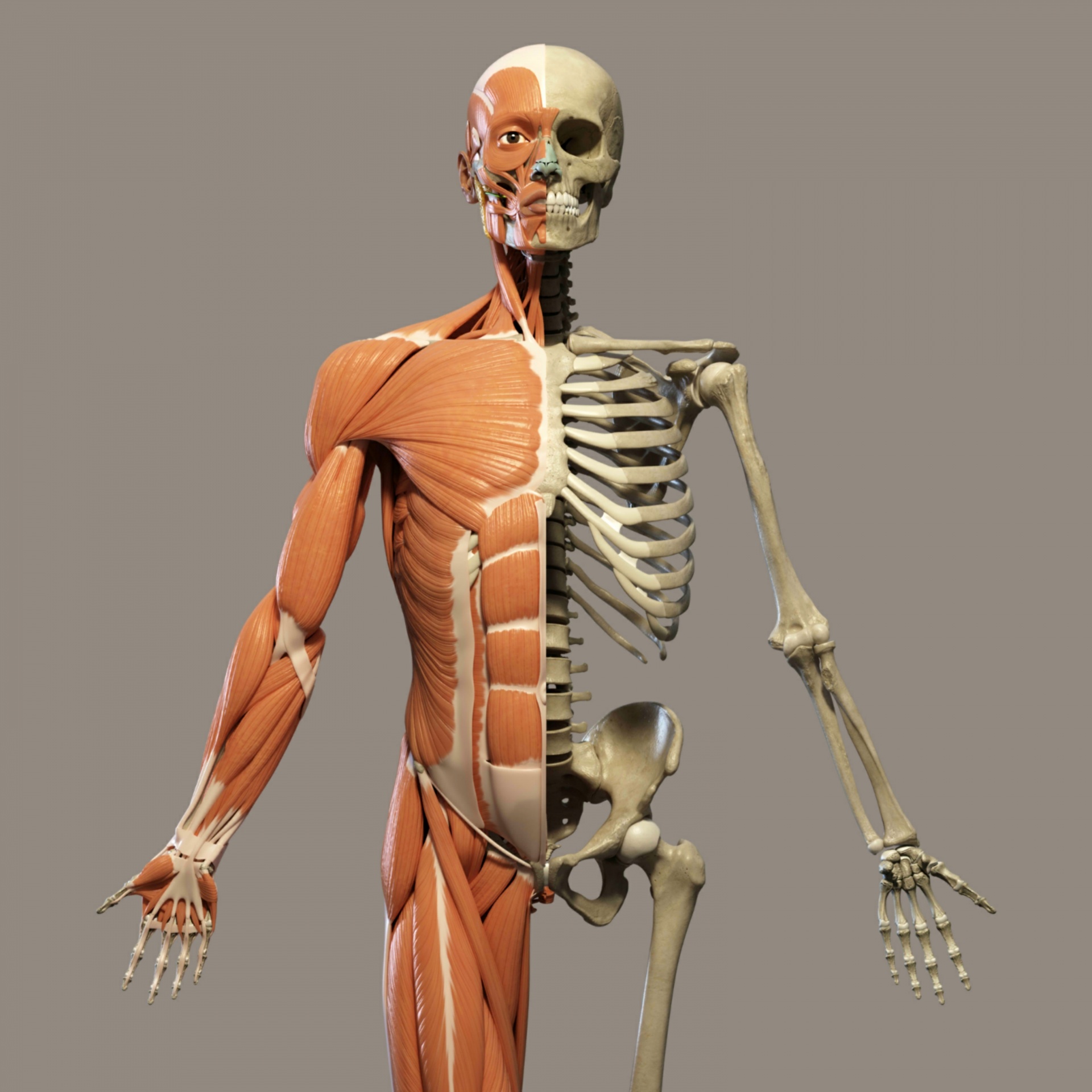
The musculoskeletal system gives your body support, stability, and movement. It is made up of the bones of the skeleton, muscles, cartilage, tendons, ligaments, joints, and other connective tissue that supports and binds tissues and organs together.
Your brain tells your body parts what to do. The muscles and bones have to work together. Muscles tend to be lonely, and don’t work by themselves, so they always come in a pair.
Muscles can only pull, they cannot push. The biceps (the top muscle on your arm) pull the arm up and the triceps (the underneath muscle) pull the arm down. Muscles are like rubber bands, they can bend, stretch and get shorter. As one muscle contracts (gets smaller) the other muscle relaxes (gets longer and looser) so that the joint (where two bones meet) can move.
The bones in your skeleton do not rub against each other when they meet at a joint, they have a special cushion in between them so we can bend without pain. The scientific, name for this cushion is cartilage.
Try the cushions activity in the Teachers' Notes page to get an idea about the importance of cartilage.
Babies have very soft bones which helps them to be born more easily. As we grow up our bones become harder and stronger. We need special vitamins and minerals to make sure they do this and the mineral calcium is important for strong bones.
But what about granny’s skeleton?
As adults get older their bones;
- become weaker.
- lose some of their very important calcium.
- have less cushioning (cartilage) between then.
This means when joints move they can be very painful and become inflamed (swollen).

In the x-ray above, the picture on the left is a normal hip joint, and on the right is an old damaged one.
Having a hip replacement is the most common solution to this kind of pain. Do you know anyone who has an artificial hip?